Mixed Infection in Microsporidial Keratitis In A Rural Tertiary Care Hospital
Shilpa K1, Venkataramana A1, Nandini C1
Shilpa K1, Venkataramana A1, Nandini C1,
Department of Ocular Microbiolog, Narayana nethralaya, Bangalore
- *Corresponding Author:
- Shilpa K
Department of Ocular Microbiology, Narayana nethralaya, Bangalore. india
Tel: 7892110106
E-mail:shilpamicro.kmc@gmail.com
Received Date: June 16, 2021; Accepted Date: October 7, 2021; Published Date: October 18, 2021
Citation: Shilpa K, Venkataramana A, Nandini C (2021 Mixed Infection In Microsporidial Keratitis In A Rural Tertiary Care Hospital. J Med Mycol Open Access Vol:7 No:5.
Abstract
Aim
To study the prevalence of Mixed infection in Microsporidial keratitis infection from a rural tertiary care hospital.
Method
Corneal scraping samples from October 2018 to October 2019 had come to Microbiology central laboratory at Narayana nethralaya hospital,Bommasandra Bangalore. 94 samples were collected using a surgical scalpel blade no. 15 and it was subjected to microscopic Gram stain examination, 10%Pottasium hydroxide (KOH), calcoflour mount and culture techniques. 16 samples showed microsporidia in KOH and Calcoflour mount, 3 samples showed bacteria in Gram stain along with microsporidia out of which, 1 Gram negative bacilli and 2 Gram positive cocci. Gram positive cocci showed growth on blood and chocolate agar after 48 hours of incubation at 37ºC, Gram negative bacilli did not showed any growth. Gram positive cocci was identified as Staphylococcus epidermidis by catalase and coagulase biochemical tests.
Results
Out of 94 samples, 16 samples showed microsporidia in KOH and Calcoflour mount, 3 samples showed bacteria in Gram stain along with microsporidia out of which, 1 Gram negative bacilli and 2 Gram positive cocci. Gram positive cocci showed growth on blood and chocolate agar after 48 hours of incubation at 37ºC, Gram negative bacilli did not showed any growth. Gram positive cocci was identified as Staphylococcus epidermidis by catalase and coagulase biochemical tests.
Conclusion
Out of 16 microsporidial cases three samples were mixed with Gram positive and Gram negative bacteria, Gram negative mixed infection was seen in above 50 years age group and Gram positive mixed infection was seen in 30-45 years age group.
Keywords
KOH, Calcoflour, Gram stain, Mixed infection.
Introduction
Microsporidia constitute a group of obligate intracellular organisms encompassing several genera. These small, oval, eukaryotic intracellular spore-forming protozoan parasites are widely distributed in vertebrates and invertebrates. Microsporidial keratitis was first reported in 1973 and was initially thought to be associated with immunocompromised hosts. Awareness of microsporidial keratitis has increased in recent years and a few cases have been reported, especially in tropical and semitropical countries[1].
Routine diagnosis of microsporidia in clinical laboratories has relied mostly on special staining and microscopic techniques. Although it has been reported that the microscopic evaluation of corneal scrapings using different stains for the diagnosis of microsporidial keratitis is sensitive, these often require expertise and the inability to differentiate related species in a specimen may require newer methods to be introduced for the identi?cation of microsporidia causing keratitis. Accurate identi?cation of microsporidia is vital for making a quick diagnosis[2]. The transmission may involve person to person as well as waterborne or food contamination, especially in developing countries with poor sanitation [3].
Materials and methods
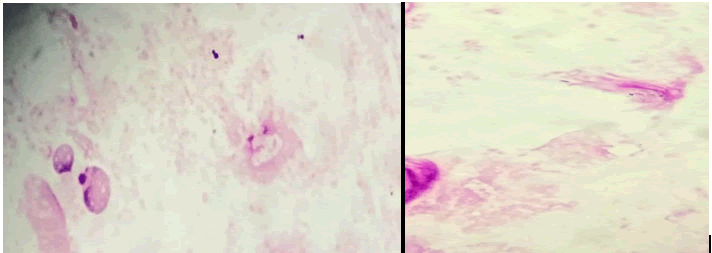
Corneal scrapings samples of the 94 patient were collected using a surgical scalpel blade no. 15 and it was subjected to microscopic Gram stain examination, 10%Pottasium hydroxide (KOH) mount, Avistain Calcoflour white stain, Modified acid fast stain (1% sulphuric acid as the decoloriser ) and culture techniques. 16 samples showed oval shaped bright apple green fluorescence microsporidia spores measuring 0.9-1.5µm to 1.2-2µm in KOH-Calcoflour mount when observed with fluorescent microscope 40 x objective. 3 samples showed bacteria in Gram stain along with Gram positive oval, bilateral shaped microsporidia under oil immersion 100x objecctive. out of which, 1 Gram negative bacilli and 2 Gram positive cocci. Gram positive cocci showed growth on blood and chocolate agar after 48 hours of incubation at 37ºC, Gram negative bacilli did not showed any growth. Gram positive cocci was as Staphylococcus epidermidis by catalase and coagulase biochemical tests. Modified acid fast stain (1% sulphuric acid as the decoloriser), shows acid fast oval microsporidial spores.
Results
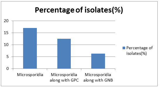
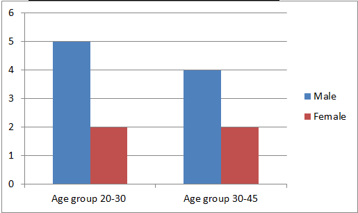
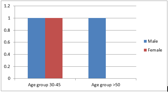
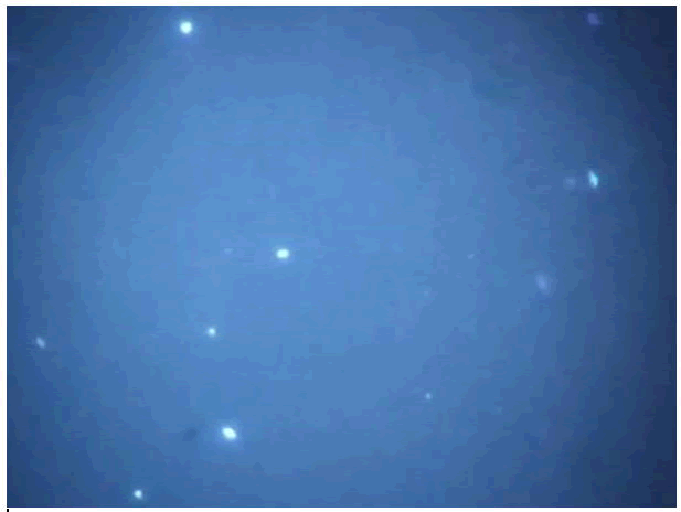
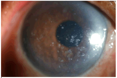
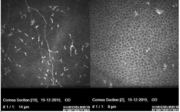
On microscopic examination, 16 samples on KOH and Calcoflour white mount reveals microsporidia(17.2%)(Fig:b). Out of which three samples showed microsporidia along with Gram positive cocci (12.5%) and Gram negative bacilli (6.25%)(Fig:a) (Table:1, Fig.1), which indicates an mixed infection of microsporidial keratitis. Seven cases of microsporidia in 20-30 age group and six cases in 30-45 age group are seen (Table:2, Fig:2). Two cases of Gram positive cocci mixed infection is seen in 30-45 age group and one case of Gram negative bacilli is seen in above 50 age group (Table:3, Fig.3). Clinical findings reveals Microsporidial keratoconjunctivitis which includes follicular / papillary conjunctivitis with multifocal, coarse corneal epitheliopathy and Confocal microscopy shows multiple rosette-like clusters / plaque of hyper reflective epithelial cells, with hyper reflective small oval intracellular (intra epithelial / intra keratocyte) bodies (Fig:c and d)
| Percentage of isolates (%) | |
|---|---|
| Microsporidia | 17.02 |
| Microsporidia along with Gram positive cocci | 12.5 |
| Microsporidia along with Gram negative bacilli | 6.25 |
Table1: Percentage of Micrsporidia and mixed infection.
| Sex | Age group 20-30 | Age group 30-45 |
|---|---|---|
| Male | 5 | 4 |
| Female | 2 | 2 |
Table2: Age and gender wise distribution of Microsporidia.
| Sex | Age group 30-45 | Age group >50 |
|---|---|---|
| Male | 1 | 1 |
| Female | 1 | 0 |
Table3: Age and gender wise distribution of Mixed infection.
Discussion
Microsporidia are now well recognised as opportunistic infectious pathogens in patients with AIDS. Diffuse superficial keratopathy and conjunctivitis are the usual clinical presentations of ocular microsporidiosis in these AIDS patients. But the clinical presentation of ocular microsporidiosis is different in immunocompetent patients[4]. Microsporidial infections in the immunocompetent patient may result in self-cure after mild symptoms appear. Con?rmation of microsporidial conjunctivitis or keratitis can be made from the examination of conjunctival or corneal swab, scraping, or biopsy specimens[5].In our retrospective study of microsporidial keratitis infection, we found 16 samples on KOH and Calcoflour white mount reveals microsporidia (17.2%). Out of which three samples showed microsporidia along with Gram positive cocci (12.5%) and Gram negative bacilli (6.25%), which indicates an mixed infection of microsporidial keratitis. Modified acid fast stain (1% sulphuric acid as the decoloriser) shows acid fast oval microsporidial spores. Clinical findings shows microsporidial keratoconjunctivitis which includes follicular/papillary conjunctivitis with multifocal, coarse corneal epitheliopathy, Confocal microscopy examination reveals multiple rosette-like clusters / plaque of hyper reflective epithelial cells, with hyper reflective small oval intracellular (intra epithelial / intra keratocyte) bodies. Seven cases of microsporidia in 20-30 age group and six cases in 30-45 age group are seen. Two cases of Gram positive cocci mixed infection is seen in 30-45 age group and one case of Gram negative bacilli is seen in above 50 age group.
The spore is the infectious stage of the microsporidian life cycle stage and the only stage that can survive outside the host cell. The shape of the microsporidian spore is usually pyriform or oval, and the spores vary in size from approximately 1 to 12μm, although some needle-like spores can be as long as 20μm[6]. Microsporidial spores were confirmed by smear examination and they do not grow in conventional culture media. The lower rate of positive culture also might be due to culture of corneal scraping in all cases irrespective of prior medication and size[7]. Both Gram-positive and Gram-negative bacterial infections may coexist (mixed infections) and are, common due to excess amounts of gut-derived LPS released into the bloodstream in several clinical conditions that alter the mucosal permeability of the intestines or due to sepsis-associated intestinal hypoperfusion[8]. Maintenance of established microsporidia cultures is relatively simple. The cultures should be examined microscopically before replenishing the medium or subculturing them. In well established cultures, the medium may often look cloudy and appear contaminated. Microscopic examination, preferably with the use of an inverted microscope equipped with differential interference contrast or phase optics[9]. Microsporidial keratitis is a rare condition with poor medical response and no treatment guidelines. Almost all reported cases of microsporidial stromal keratitis failed medical therapy and required penetrating keratoplasty[10].
Conclusion
From our retrospective study we would like to conclude that microsporidial keratitis is a rare condition in which there is a mixed infection of Gram positive and Gram negative bacteria in 30 to above 50 years of age group in which males were more affected than females.
References
- Satoru Ueno, Hiroshi Eguchi, Fumika Hotta, Masahiko Fukuda, Masatomo Kimura, Kenji Yagita. Microsporidial keratitis retrospectively diagnosed byultrastructural study offormalin-fixed paraffin-embedded corneal tissue.Ann Clin Microbiol Antimicrobials. 2019; 18(17): 1-5.
- Joveeta Joseph,Savitri Sharma,Somasheila Murthy, Pravin V. Krishna,Prashant Garg, Rishita Nutheti . Microsporidial Keratitis in India: 16S rRNA Gene-Based PCR Assay for Diagnosis and Species Identiï¬Âcation of Microsporidia in Clinical Samples. Invest Ophthalmol. 2006; 47(10): 104468-4473.
- Rajesh Fogla, Prema Padmanabhan, K Lily Therese, Jyotirmay Biswas, Madhavan. Chronic Microsporidial Stromal Keratitis in an Immunocompetent, Non-Contact Lens Wearer. Indian J Ophthalmol. 2005; 53(2): 123-125.
- LSG & Associates, Santa Monica. Laboratory Identiï¬Âcation of the Microsporidia. JOURNAL OF CLINICAL MICROBIOLOGY.2002;40(6):1892–1901.
- Bing Han and Louis M. Weiss. Microsporidia: Obligate Intracellular Pathogens within the fungal kingdom. Microbiol Spectr.2017; 5(2): 1-26.
- Sujata Das, Ruchipriya Samantaray, Aparajita Mallick, Srikant K Sahu, Savitri Sharma. Types of organisms and inâ??vitro susceptibility of bacterial isolates from patients with microbial keratitis: A trend analysis of 8 years.2018; 67(1): 50-53.
- Pramod Jagtap,Rohini Singh, Karuna Deepika,Venkataraman Sritharan, Shalini Gupta. A Flow through Assay for Rapid Bedside Stratiï¬Âcation of Bloodstream Bacterial Infection in Critically ill Patients: a Pilot Study. J Clin Microbiol. 2018; 56(9): 1-13.
- Govinda S. Visvesvara. In Vitro Cultivation of Microsporidia of Clinical Importance. Clin Microbiol review. 2002; 15(3): 401–413.
- 10.Mircea coca,James kim,Sudhir shenoy,Patricia chevez-Barrios,Manuj Kapur. Microsporidial Stromal Keratitis: Successful Treatment with Topical Voriconazole and Oral Itraconazole. Cureus.2016; 8(12): 9-34.
Open Access Journals
- Aquaculture & Veterinary Science
- Chemistry & Chemical Sciences
- Clinical Sciences
- Engineering
- General Science
- Genetics & Molecular Biology
- Health Care & Nursing
- Immunology & Microbiology
- Materials Science
- Mathematics & Physics
- Medical Sciences
- Neurology & Psychiatry
- Oncology & Cancer Science
- Pharmaceutical Sciences
